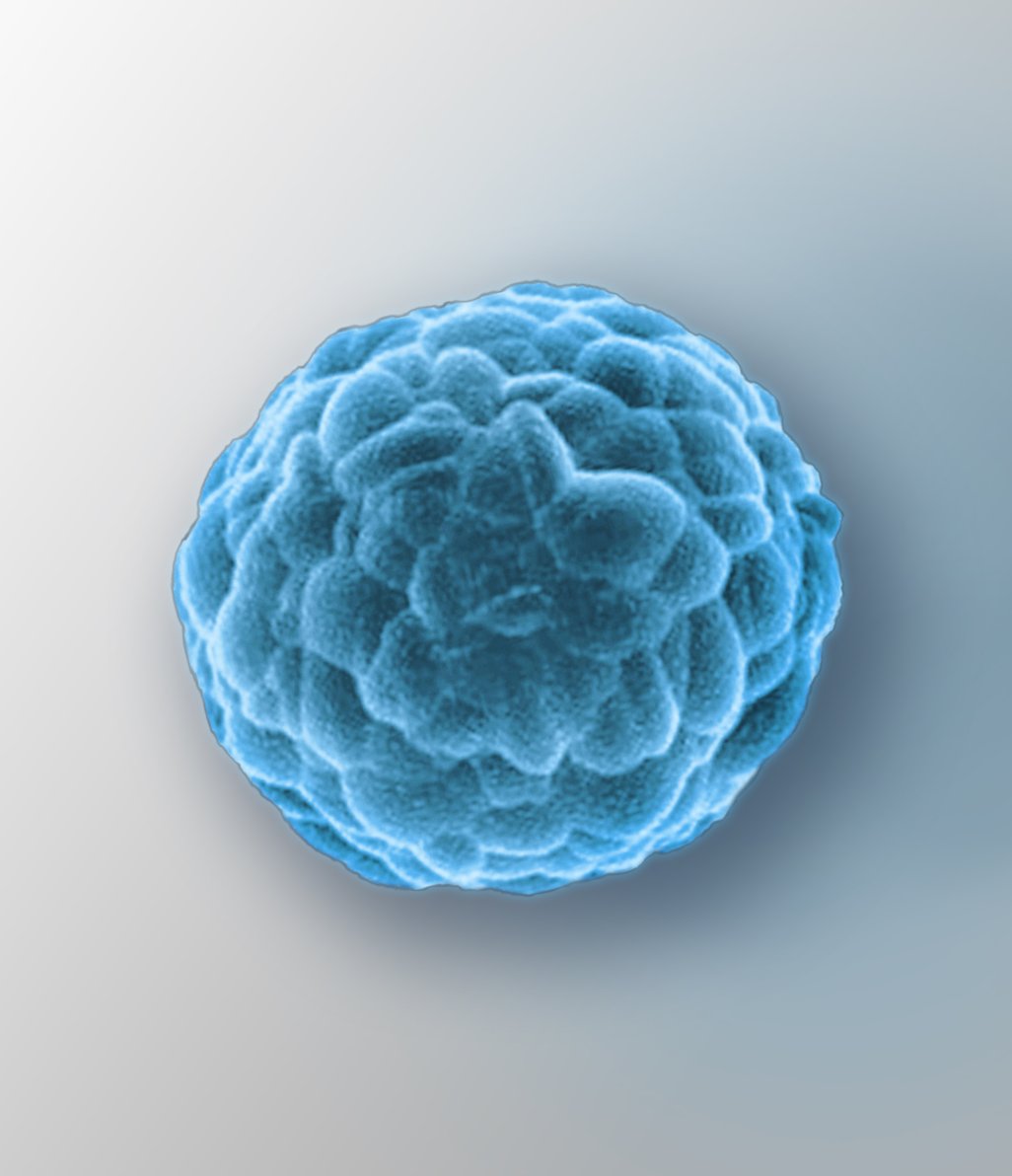
Take a closer look. See results.
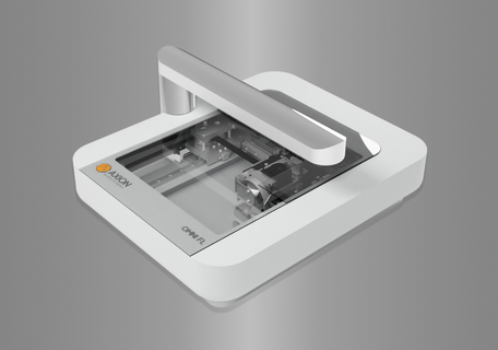
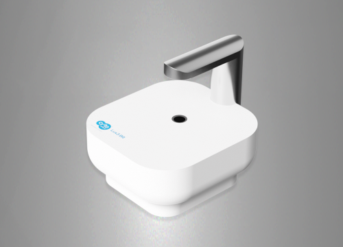
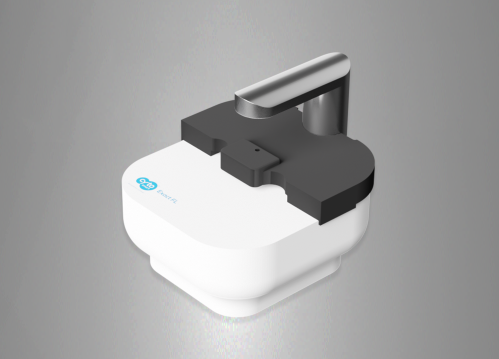
Image your cells in the incubator and get a clearer picture of your biology with our live-cell analysis platforms.


.png")
Image your cells in the incubator and get a clearer picture of your biology with our live-cell analysis platforms.