Authors: Buesch S, Schaepermeier S, D'Souza T, Ortmann B, Schwartz C, Schroeder J.
Lonza Bioscience Solutions, 2017
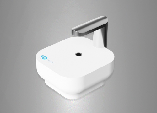
Movement of cells plays a critical role in the development of cancer. Analyzing the motility of cells in appropriate cell culture models is therefore an important tool for cancer researchers. Live cell imaging is particularly well suited to capture dynamic processes in cell culture. The CytoSMART™ Device is an easy-to-use, small and affordable live cell monitoring system suitable for the label-free analysis of cell motility within a standard cell culture incubator. Label-free approaches offer the benefit of cell analysis without potential cytotoxic or other side-effects of the used markers or dyes on the cells. This presentation shows the suitability of the CytoSMART™ Device for the analysis of different cancer-relevant assays. The formation of new blood vessels is required to ensure sufficient nutrient and oxygen supply and to allow solid tumors to grow beyond a certain size. This process can be mimicked in cell culture models in so-called tube formation assays. In this study, Human Umbilical Vein Endothelial Cells (HUVEC) were seeded on Engelbreth-Holm Swarm Sarcoma derived Basement Membrane Extract (BME). The resulting formation of endothelial tubes was monitored with the CytoSMART™ Device. Subsequently, the average length of the formed tubes as well as the number of closed tube circles was quantitatively determined. In addition, the impact of Suramin on tube formation was evaluated as an example for a tube formation inhibiting compound. The migration of cancer cells is also required for the growth and in particular the metastasis of tumors. In a first example, the closure of a so-called wound or scratch in a confluent monolayer of cancer cells was monitored with the CytoSMART™ Device. Determining the migration speed of the cells by measuring the speed of wound closure is a simple assay to determine the migration potential of cancer cells. Modifying the cancer cells, e.g. by knocking down specific genes with siRNA, can help to identify genes that play a role in cell migration. Compounds that are expected to reduce cell motility and therefore reduce the metastasis potential of cancer cells can be easily tested. In a second example, the invasion of cancer cells into a three-dimensional (3D) matrix was analyzed. Tumor cells were embedded in a cancer-relevant matrix and their invasion into the 3D matrix was documented with the CytoSMART™ Device. While this type of model is slightly more difficult in set-up and analysis compared to simple scratch assays, it may reflect better the in vivo situation where solid tumors develop within three-dimensional tissues. The images captured with the CytoSMART™ System were quantitatively analyzed using appropriate software. Overall, the CytoSMART™ System is an easy-to-use, small and affordable live cell imaging system suitable for the label-free analysis of different cancer-relevant assays.