Accelerate your research with fluorescence cell counting
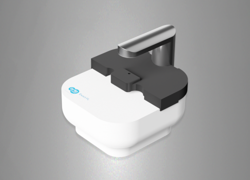
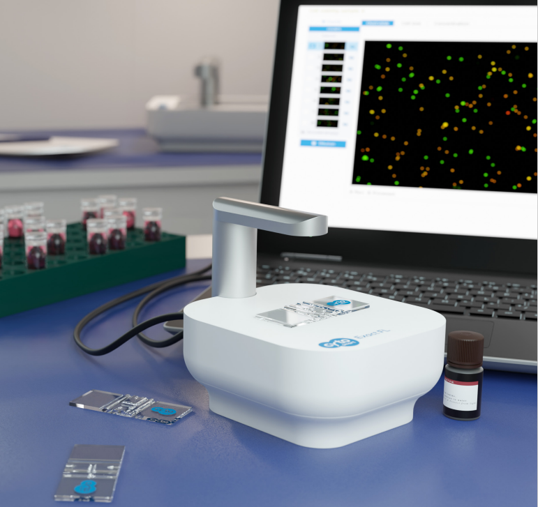
Automated fluorescence cell counting has transformed the process of cell quantification, as it offers a great deal of flexibility and accuracy, and can be used when bright-field image analysis is not possible. The CytoSMART Exact FL is an automated, dual fluorescence cell counter with an expanded field of view and unmatched resolution and magnification power. Using an advanced optical system and AI-powered image analysis software, the CytoSMART Exact FL calculates the exact number of cells in a sample and provides reliable assessment of key cellular parameters, including cell viability and transfection efficiency.
The CytoSMART Exact FL features:
- >> State-of-the-art optics allows to accurately distinguish individual cells in clumps and count cells down to 4 µm in diameter
- >> Dual-channel fluorescence can be used for examining health and viability of hard-to-detect-cells
- >> AI-powered software performs cell counting, minimizing user-to-user variability
- >> Large field of view & multi-count let users analyze a larger portion of the available counting volume
- >> Add-ons (e.g. organoid counting) expand the application range of the device
- >> Reusable or disposable counting slides for cost- or time-efficient cell counting
Applications
The use of fluorescence cell counting significantly accelerates experimental work in the fields of cancer biology, immunology, tissue engineering, and cell therapy. Numerous cell-based assays significantly benefit from fluorescence cell counting due to the number of issues associated with detecting and analyzing mammalian cells using colorimetric dyes (e.g. Trypan Blue). The CytoSMART Exact FL, fitted with red and green fluorescent channels, in combination with appropriate fluorescent dyes, can assist you in examining the following features (Fig. 1):
- >> Cell viability
- >> Small and hard-to-detect cells
- >> Primary cell samples
- >> Heterogeneous cell populations
- >> Transfection efficiency
- >> Distinguishing whole cells from debris
- >> and more.
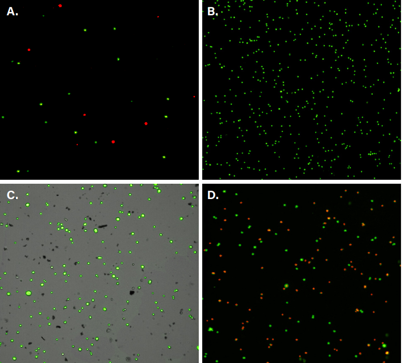
Figure 1: Examples of applications of the Exact FL Fluorescence Cell Counter. (A) Examining macrophage subpopulations. Polarized M1 macrophages were expressing a red fluorescent protein (RFP), whereas M2 macrophages were tagged using a green fluorescent protein (GFP), allowing to visualize and distinguish between two distinct macrophage subgroups. (B) Counting peripheral blood mononuclear cells (PBMCs). The cells were treated with acridine orange (AO) that emits green fluorescence when bound to double stranded DNA. (C) Distinguishing between C6 rat glioma cells and cellular debris. AO was applied to C6 cell samples to count the total number of cells. (D) Assessing viability of 3T3 mouse embryonic fibroblasts. The cells were stained with AO and propidium iodide (PI), allowing to access the proportion of alive (green) and dead (red) cells in a sample.
State-of-the-art optics
Using the 6.4 MP CMOS camera combined with 10× magnification, the CytoSMART Exact FL can visualize and count cells down to 4 µm in diameter. With increased field of view and multi-count feature, it is also possible to take multiple snapshots of the same sample at various slide positions, which increases the accuracy and reproducibility of a cell count.
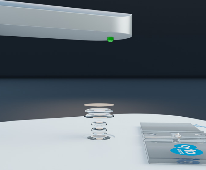
Figure 2: The advanced optical system of the CytoSMART Exact FL allows to visualize a range of cells (4 – 70 µm).
AI-powered algorithms
Manual cell counting introduces user-to-user variability to a scientific study. In addition, the process of manual cell counting is challenging and time-consuming, especially when working with cells that are prone to clumping. Equipped with a robust pattern recognition software and a declustering algorithm, the CytoSMART Exact FL detects resuspended single cells, as well as individual cells within cellular clusters with high accuracy and without any bias.
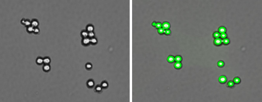
Figure 3: Raw data (left) is processed by the highly trained deep-learning neural network algorithm (right) that can analyze even the most challenging samples.
Freedom to choose between reusable or disposable counting slides
The CytoSMART Exact FL is compatible with reusable counting chambers, which makes the process of automated cell counting much more cost-effective. Customers that prefer working with disposable slides, can also use the Exact FL with a range of disposable cell counting chambers.
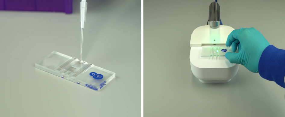
Figure 4: The CytoSMART Exact FL is compatible with a range of glass (left) or plastic slides, as long as the chamber depth does not exceed 0.1 mm (cell counting) and 0.2 mm (organoid counting). In addition, the device is equipped with a plastic stage cover (right) to prevent environmental light from having an effect on fluorescence cell counting.
Automated software updates & secure cloud data storage
All CytoSMART devices are connected to the CytoSMART cloud environment. This means that the data generated by the Exact FL fluorescence cell counter is processed, analysed and stored in one accessible and secure place, protected by a multi-layered cyber security system. Novel software updates are also introduced via the CytoSMART Cloud. With only a few clicks our customers can download and install these add-ons to make their user experience even more seamless. Importantly, the added features will not affect consistency and reproducibility of acquired results.
Innovative organoid counting software
Organoids are stem cell-derived 3D culture systems that have been used to study genetic disorders, infectious diseases, and various types of cancer. To ensure consistency and reproducibility across multiple organoid-based experiments, it is critical to use a fixed concentration of organoids of similar size. Using the Organoid Counting add-on, the CytoSMART Exact FL can analyse bright-field images to determine precise number and size of organoids in a sample.
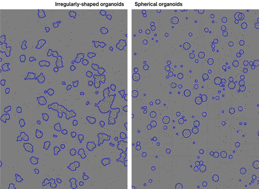
Figure 6: The newly updated Organoid Counting algorithm is designed to recognize and quantify not only irregularly-shaped organoids (left), but also spherical organoids (right). In addition, the new algorithm is better at differentiating organoids from cellular debris, thus reducing the amount of false positives/false negatives.
Frequently Asked Questions
Q: Which fluorescent dyes can be used with the CytoSMART Exact FL?
A: Many different fluorescent dyes can be used, as long as the fluorescent dye’s excitation and emission spectra correspond with the fluorescent filters of the Exact FL (green – excitation: 452/45 nm, emission: 512/23 nm; red – excitation: 561/14 nm, emission: 630/90 nm). Some examples are propidium iodide (PI) and red fluorescent protein (RFP) for the red channel, and acridine orange (AO), calcein-AM, and green fluorescent protein (GFP) for the green channel.
Q: Can the CytoSMART Exact FL access cell viability?
A: Yes, the viability of cells can be assessed either using the green/red fluorescent channels (AO/PI) or a bright-field channel (Trypan Blue).
Q: Is a computer required?
A: The device requires a dedicated desktop or laptop running on Windows 10 or above and with a USB3 port and an active internet connection. A WiFi or Ethernet connection is needed for connecting to the CytoSMART Cloud for image analysis and storage.
Q: Do I need to calibrate the CytoSMART Exact FL?
A: The calibration is not required.
Q: Is it required to use a light cover when performing fluorescence cell counting?
A: To get the best results, we advise covering the stage of the device with the provided stage cover to minimize the amount of environmental light.
Q: Is it possible to control the intensity of the Exact FL LED?
A: Yes, it is possible to set the intensity of the LED for red and green fluorescent channels according to users’ preferences.
Q: Can I clean the CytoSMART Exact FL?
A: The device is easy to clean using lint-free wipes and ethanol (70%) or isopropyl alcohol (IPA). Do not use acetone to clean the device, also the device cannot be autoclaved.
Technical Specifications
Ordering Information
Request quote here.