Brightfield imaging (or brightfield microscopy) involves transmission of illumination light through the sample, with the contrast being generated as a result of the light absorption by dense parts of the specimen. As opposed to fluorescence microscopy, brightfield microscopy only utilizes white light for sample illumination. The sample doesn’t need to be labelled or modified in any way, which allows to preserve cells in their native, undisturbed state and also saves time and resources. Brightfield live-cell imaging has become an increasingly valuable tool in cell research to monitor, quantify, and analyze proliferation and activity of cells, specifically cell motility, apoptosis, and cell differentiation.
CytoSMART Technologies offers a range of brightfield live-cell imagers, adapted to varying applications and budgets. Our devices are designed to function inside a cell culture incubator, ensuring that cells remain in their desired culturing environment throughout the whole imaging period. The utilized digital phase contrast technique produces high-contrast, publication-quality images. The devices are equipped with artificial intelligence (AI)-driven image analysis algorithms (e.g. cell confluency, colony detection, cell migration) that process the acquired images and generate automatic reports. These reports contain detailed information on multiple analyzed variables and can be assessed remotely via the CytoSMART Cloud.

CytoSMART Omni
The CytoSMART Omni is a brightfield microscope that can visualize an entire surface area of a culture vessel by automatically acquiring multiple images and stitching them. The 6.4 MP CMOS camera moves along the sample platform, without disturbing the sample.
Learn more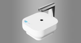
CytoSMART Lux3 BR
The CytoSMART Lux3 BR is an inverted digital microscope, which utilizes brightfield imaging with digital phase contrast to acquire publication-quality images and videos of cells.
Learn more